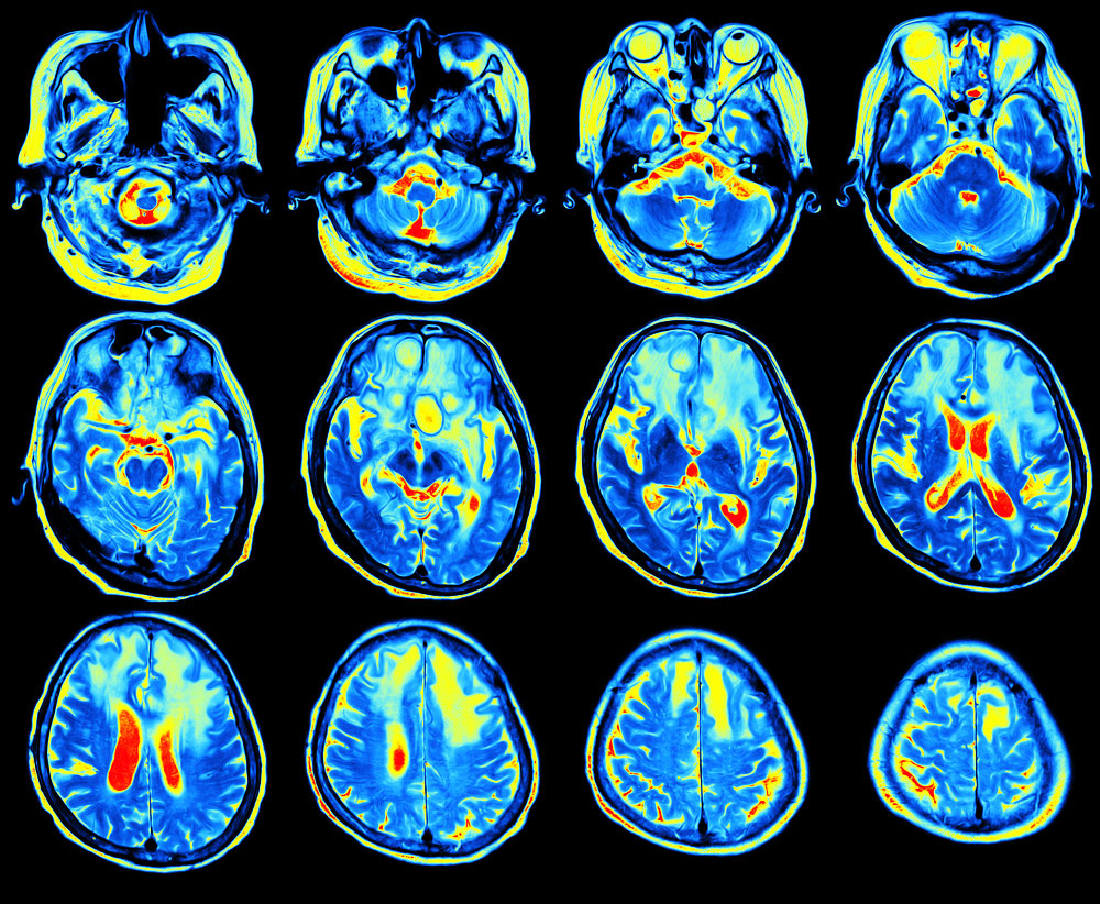
centers in the brain. The factors determining risk during the monetary incentive task were mainly found in the anterior region of the putamen with overlapping portions of caudate, nucleus accumbens, and globus pallidus. The part of the brain that displayed activity during the go/no-go task was the anterior insula bilaterally, with other strong activity in right inferior frontal gyrus, putamen, and thalamus. Finally, the evocative images task showed activity in the visual cortex and the amygdala bilaterally.
“[The fMRI] offers what we call an intermediate phenotype — a measure of brain activity that is a proxy for predictions of addiction — but which may be more reliable and also closer to mechanisms that can be interrupted by medicines,” Nutt said. “If we can turn off an overactive system in the fMRI with a drug, this might encourage a trial of this drug in that patient. We have a long way to validate this approach as we need to take drugs that are promising in the fMRI into clinical trials. But the fMRI approach is quicker and cheaper than clinical trials so more drugs can be screened.”
Nutt and his team of researchers aimed to establish a platform and framework for further studies to better determine how these centers in the brain affect addiction.
“It could help us find new treatments,” Nutt said. “And also possibly select patient groups that might respond best to certain medicines.”


